 Synedra acus
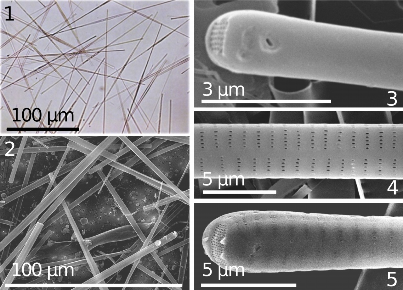
 subsp.
radians. Figs. 1-2: Cells. Figs 3, 5: Valve ends with rimoportulae. Fig. 4: Central area. LM – Fig. 1; SEM – Figs 2-5. Magnification: Fig. 1 – ×110. Courtesy of Ye. V. Likhoshway.
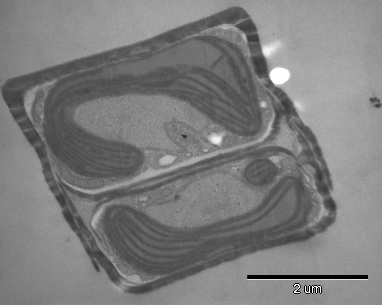
Dividing cell of
Synedra acus subsp.
radians, ultra-thin transverse section, TEM. Courtesy of Ye. D. Bedoshvili.
 Synedra acus
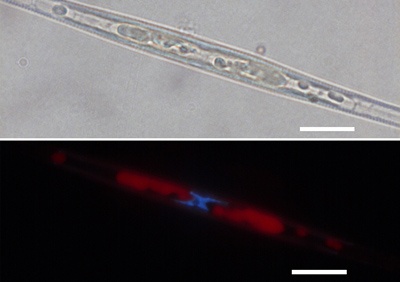
 subsp.
radians, cells fixed with 70% ethanol followed by DAPI staining. Light microscopy. Above – white light, below – fluorescence of chloroplast (red) and nucleus (blue). Scale bar = 10 μm. Courtesy of Ye. D. Bedoshvili.
 Synedra acus
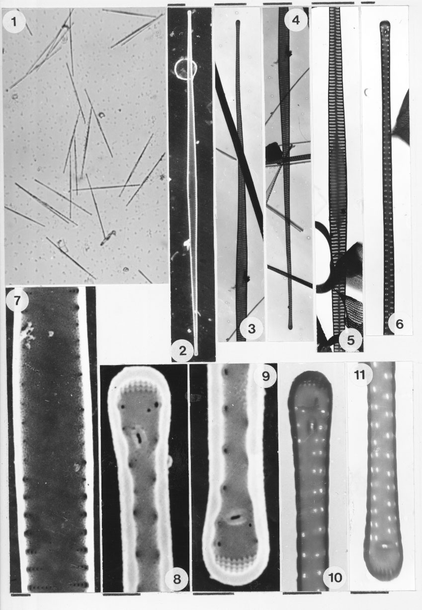
 subsp.
radians. Fig. 1: Cells. Fig. 2: Valve internal surface. Figs 3, 4: Valve ends. Fig. 5: Central area. Fig. 6: Opening of rimoportula at valve end. Figs 7-9: Details of external valve surface. Fig. 7: Central area. Figs 8-11: Valve ends with rimoportulae. LM – Fig. 1; SEM – Figs 2, 7-9; TEM – Figs 3-6, 10-11. Magnification: Fig. 1 – ×110. Scale bars = 10 µm (Figs 2-5); 1 µm (Figs 6-11). Courtesy of Ye. V. Likhoshway.

Whole live cell of
Synedra acus subsp.
radians, light microscopy. Scale bar = 10 μm. Courtesy of Ye. D. Bedoshvili.

Whole valve of
Synedra acus subsp.
radians, SEM. Courtesy of A. D. Firsova.

